A medical imaging device that utilizes sound waves to produce images, the ultrasound is a valuable diagnostic tool. Sound waves bounce off of the various internal structures and substances – tissue, bone, blood and amniotic fluid, for example – with varying rates and speeds of return. That data is collected, then assembled into an image via computer. Because it doesn’t use radiation, it is considered safe for use during pregnancy to track fetal development. However, that is far from its only use.
Ultrasounds are used to obtain visual information about internal organs and systems, including the cardiovascular and digestive systems. This type of medical imaging device can also be used to provide a visual guide for such internal procedures as obtaining a sample for biopsy. The type of equipment used and the complexity of the image can vary according to the specific ultrasound procedure. Quite simply, the ultrasound is one of the most powerful and dynamic medical diagnostic tools available today.
When Do You Need an Ultrasound?
Ultrasounds have become a standard part of pregnancy care. The first is usually early in the pregnancy, checking the development of the heart and determining the due date. The second is around the 20-week mark, checking fetal development. Depending on the circumstances, there may be more ultrasounds required during a pregnancy, such as if the mother is at high risk of preterm labor.
An ultrasound is also used when a body exhibits symptoms that require an internal view to make a diagnosis. Abdominal pain and swelling may require an ultrasound to check the organs in that area, check for blockages and internal irregularities, such as tumors or cysts. An ultrasound can be used to check heart function and blood flow through the cardiovascular system. Ultrasounds can also be used as a visual aid in internal procedures and treatments, including tumor treatments and biopsies.
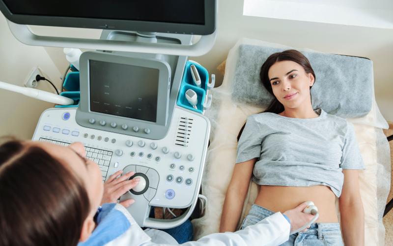
The Ultrasound Process
Depending on the type of ultrasound and the part of the body undergoing the procedure, there may be some preparation involved. For some types of ultrasounds, the bladder may need to be full. For others, a pre-ultrasound fast may be required. This helps to ensure that the desired internal area of the body can be viewed as accurately as possible. Your health care provider will let you know exactly what is required prior to the procedure.
At the time of the ultrasound, the skin over the procedure area will be exposed, then spread with a non-staining gel. The gel makes it easy to move the transducer over the ultrasound area and helps with sound wave transmission. The transducer produces sound waves at a pitch outside of human hearing range. Some ultrasound procedures require the insertion of a transducer. These include the transvaginal and the transrectal ultrasounds. Ultrasounds usually take about 30 to 45 minutes, depending on the complexity of the situation or the amount of information to be gathered. After image analysis, your healthcare provider will discuss the results with you.
Excellent Diagnostic Tool
Ultrasound technology offers many benefits as a diagnostic tool. It is less invasive than the exploratory surgeries patients faced a generation ago. Today’s ultrasound offers impressive accuracy, with 3D and even 4D real time imaging available when required during pregnancy care. Using ultrasound guidance during internal medical procedures can mean smaller incisions and increased precision. As a general consideration, Ultrasounds make it easier to diagnose issues, which results in more efficient and less invasive treatment, as well as less complications. It really is a useful medical test.